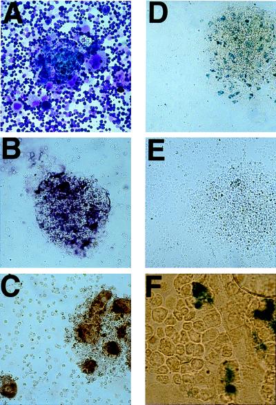
Figure 5.
Retroviral infection of TVA-expressing cells in live animals. After i.p. injection of 0.25–0.5 × 10⁶ env-A coated, β-gal-expressing infectious particles on each of 2 consecutive days, mice were killed on the next day. Wright-Giemsa staining (A) of freshly harvested bone marrow demonstrated the megakaryocytes to be mostly in clumps. Cells that stained positive for CD41 (B) and for TVA (C) were predominantly located in the megakaryocyte-containing clumps. β-gal staining demonstrated positive cells limited to the small cells within the megakaryocyte-containing clumps (D). No staining was seen in bone marrow from nontransgenic control mice subjected to the identical infections (E). A region with preserved bone marrow architecture from a transgenic mouse (F) demonstrates β-gal expressing cells adjacent to a bone marrow blood vessel.

