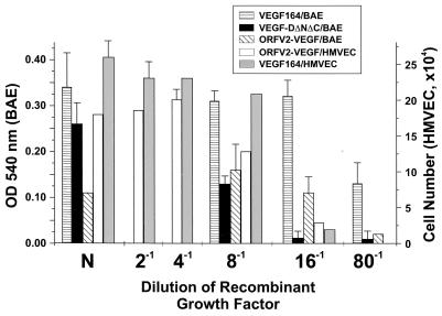
Figure 6.
Mitogenic activity of purified ORFV2-VEGF on endothelial cells. BAEs and HMVECs were treated with either purified ORFV2-VEGF, mouse VEGF164, or VEGF-DΔNΔC diluted in medium with reduced serum and without added growth factors (in the case of HMVEC). The factors were used either neat (N) or diluted 1:2, 1:4, 1:8, 1:16, or 1:80 prior to use. The neat concentration of factor was (VEGF164/BAE, 80 ng/ml; VEGF-DΔNΔC/BAE 80 ng/ml; ORFV2-VEGF/BAE, 80 ng/ml; ORFV2-VEGF/HMVEC 400 ng/ml; and VEGF164/HMVEC, 200 ng/ml). Note that the BAE have been tested at N, 1:8, 1:16, and 1:80 and the HMVEC cells at N, 1:2, 1:4, 1:8, and 1:16 only. After 72 h, the amount of cellular proliferation was quantitated by using either MTT substrate or counting. Values expressed (mean ± standard errors, representative of three experiments) are the cell number or proliferation above that were induced by the assay medium alone.

