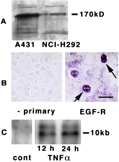
Figure 1.
Expression of EGF-R in cultured cells. (A) Immunoblotting of EGF-R in A431 and in NCI-H292 cells. Cells were examined after becoming confluent. Lysates were electrophoresed in 8% acrylamide gels and blotted with anti-EGF-R antibody. Molecular mass of marker protein is reported on the right. (B) Immunocytochemical analysis with anti-EGF-R antibody in cultures of NCI-H292 cells. At confluence, positive staining was seen in most of cells, and certain cells had more intense staining (arrows, right side). In the absence of the primary antibody, no staining was seen (left side). (Bar = 25 μm.) (C) Northern analysis of EGF-R in NCI-H292 cells was performed on total RNA extracted from confluent cultures incubated with TNFα (20 ng/ml) for 12 or 24 h. The RNA (10 μg) was electrophoresed on a formaldehyde-agarose gel, transferred to a nylon membrane, and hybridized with the 32P-labeled EGF-R cDNA probe. After hybridization, the membrane was washed and autoradiographed.

