
Figure 2.
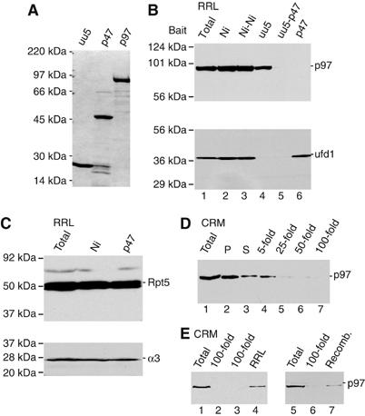
RRL depletion of p97 and p97 complexes. (A) Coomassie-stained gel of recombinant His-tagged proteins used as bait for RRL affinity depletion. (B) Immunoblots for p97 (top) and ufd1 (bottom) of intact RRL (lane 1), RRL following a single adsorption with Ni-NTA beads (lane 2), uu5-coated beads (lane 4) or p47-coated beads (lane 6), and two sequential adsorptions with Ni-NTA beads (lane 3) or uu5 followed by p47 (lane 5). (C) Immunoblots for 19S RC subunit Rpt5 (top) and 20S α3 subunit (bottom) using intact, mock- and p47-adsorbed RRL. (D) Immunoblot of membrane-associated p97. Data show p97 recovered in pellet (P) and supernatant (S) after pelleting microsomes (lanes 2 and 3) and the amount of p97 from the same starting material that remained associated with membranes after dilution indicated (lanes 4–7). (E) CRMs (lanes 1 and 5) and p97-depleted membranes (100-fold dilution) were reincubated with RRL (lane 4) or recombinant p97 (lane 7), pelleted and analyzed by immunoblotting.