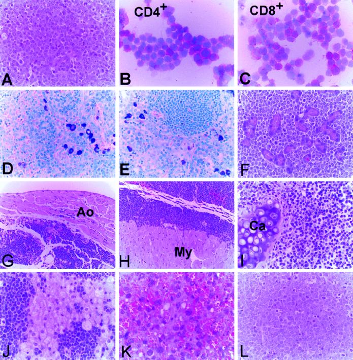
Figure 3.
Gallery of digitized images illustrating the morphology of tumors in p53/Dcn double mutant animals. Histopathological examination of the tumors revealed high-grade lymphoma (A). B and C represent cytospin preparations of the cultured PD100 lymphoma shown in A after immunostaining with anti-CD4 and anti-CD8 antibodies. The tumor cells are highly invasive and infiltrate the soft tissues of mediastinum (D and J), the salivary glands (E and F), the periaortic spaces (G), the pericardium (H), and the bronchial wall (I). K shows a section of the only nonlymphoid tumor, a high-grade hemangiosarcoma, found in a double mutant animal. L is a section of a thymic lymphoma detected in the p53−/− Dcn+/− animals showing microscopic features identical to those observed in the double mutant animals. The images were digitized with a Pixera digital camera at resolution of ≈106 pixels/inch. [×150 (A, F, I, J, L); ×300 (B, C, K); ×100 (D, E, G, H).]

