Figure 2.

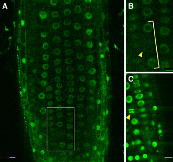
CLF protein is nuclear localised but is not present in nuclei throughout mitosis. (A) Root of a clf-50/clf-50 35S∷GFP-CLF plant showing GFP expression in most cells in the nuclei. (B) Close-up of the inset in (A) showing a cell without nuclear GFP expression (arrowhead). Adjacent cells in the same cell file show nuclear GFP (bracket). (C) Details of a VRN1∷VRN1-GFP root exhibiting a mitotic figure (arrowhead). At least 10 roots per line were analysed by confocal laser microscopy; in no case, GFP-stained mitotic figures were identified in clf/clf 35S∷GFP-CLF roots, whereas VRN1∷VRN1-GFP roots showed green-fluorescing mitotic figures in all roots. Scale bars, 10 μm.
