
Figure 2.
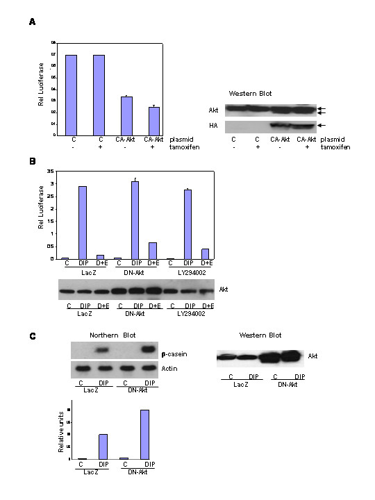
The effect of conditionally active-Akt1 and dominant negative-Akt1 on HC11 differentiation. A. The HC11-luci cells transiently transfected with either a conditionally active-Akt-1 (CA-Akt) or a control vector (pCDNA3.1). At 24 hours the cells were incubated in DIP-induction media with or without tamoxifen (1 μM). Luciferase activity was determined 48 hours post-induction and was normalized to protein concentration. Expression of Akt and HA-CA-Akt was determined via western blot. Lanes were loaded with equal amounts of protein (117.5 μg). B. The effect of dominant negative-Akt1 (DN-Akt) adenoviral infection on EGF disruption of differentiation. HC11-luci cells were infected with either a DN-Akt1 or control (LacZ) adenovirus. Cells were changed to DIP-induction media the next day and lysates were harvested 48 hours post-induction for β-casein promotor luciferase activity. The results were compared to cultures exposed to DIP plus LY294002 (5 μM). Expression of Akt was determined via western blot analysis and gels were loaded with equal amount of protein (120 μg). C: HC11 cells were infected with either a DN-Akt1 or control (LacZ) adenovirus. RNA was harvested 48 hours post-induction for analysis of β-casein RNA expression by northern blot. Expression of Akt was determined via western blot analysis and gels were loaded with equal amount of protein (120 μg).