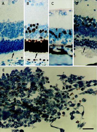
Figure 7.
FGF-2 IR in RCS rat retina. (A–D) PRC (arrows) and retinal-pigment epithelial cells (asterisks) of normal 3-month-old RCS retina (A and B) and dystrophic 111-day-old RCS rat retina (C and D). Immunostaining without primary antibody (A; control) and with anti-FGF-2 antibodies (B–D). (D) Note the lack of immunostaining in the PRC of the rescued retina. (E) Cytoplasmic FGF-2 immunostaining in encapsulated FGF18 90 days after transplantation. The sections (5 μm) were counterstained with hematoxylin. (Bar = 10 μm.)

