Figure 3.
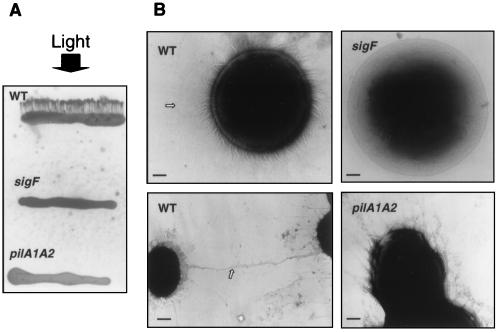
(A) Directional motility assay. Five to 10 microliters of logarithmically growing wild-type cells (WT) and the sigF and pilA1A2 mutants were streaked as an approximately 1-mm thick line onto solid (0.4% agar) BG-11 medium containing 15 mM glucose. The cells were subjected to unidirectional light (indicated by an arrow) of 40 μmol photons·m−2⋅s−1 for 48 h. The temperature during the incubation was 30°C. Note the finger-like projections emerging from the WT streak. (B) Transmission electron microscopy of whole cells. Logarithmically growing WT (Left), sigF mutant (Upper Right) and pilA1A2 mutant (Lower Right) cells. The cells were negatively stained with 1% phosphotungstic acid and examined by using a Phillips CM12 microscope. The arrow in WT (Upper Left) points to a very long pilus while the arrow in WT (Lower Left) points to pili that appear to connect neighboring cells. The bars represent 0.28 μm (Upper Left), 0.24 μm (Upper Right), 0.74 μm (Lower Left), and 0.31 μm (Lower Right).

