Figure 5.
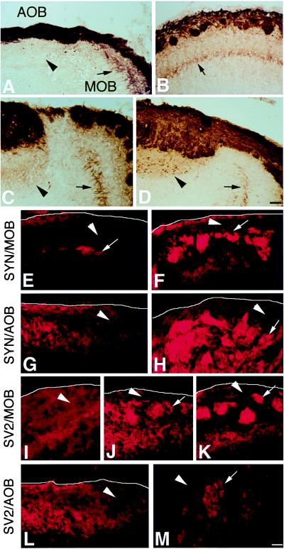
Transneuronal transfer in the AOB versus MOB during development. At birth (A), BL immunoreactivity was seen in mitral cell dendrites in the MOB (arrow), but not in the AOB (arrowhead), of PompBL mice, even though BL+ sensory axons were present in both areas. By P4, BL+ mitral cell soma (arrow) were clearly visible in the MOB (B). At P8 (C), AOB mitral cells were only faintly BL+, but became strongly BL+ by 3 weeks of age (D) (as shown in Fig. 2) (arrows and arrowheads as in A). Immunostaining of OE and VNO sensory axons in the bulb with antibodies to synaptophysin (syn) or SV2 indicated that the two proteins become localized to glomerular structures later in the AOB than in the MOB: syn in the MOB at E17.5 (E) and P0 (F), and in the AOB at P0 (G) and P8 (H); SV2 in the MOB at E15.5 (I), P0 (J), and P2 (K), and in the AOB at P0 (L) and P8 (M). In E–M, arrowheads indicate incoming sensory axons, arrows indicate axons coalescing in protoglomeruli or glomeruli, and a white line indicates the outer border of the bulb (out of the field in M). [Bar = 50 μm (A–D) and 25 μm (E–M).]

