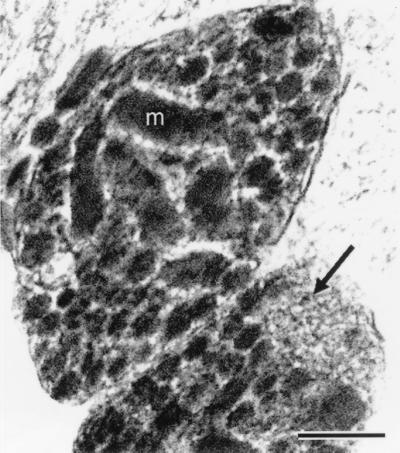
Figure 1.
Electron micrograph (low-dose, dark-field scanning transmission electron microscope (STEM) image) of a freeze-dried ultrathin (ca. 90 nm thick) cryosection prepared from a rapidly frozen pellet of isolated nerve terminals. m, mitochondrion. An arrow points to a cluster of microvesicles; large neurosecretory vesicles fill the rest of the cytoplasmic space. (Bar = 0.2 μm.)

