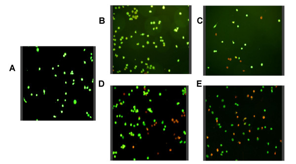
Figure 1.

Mycobacterium-induced cell death in THP-1 cells. THP-1 cells were plated on coverslips at 2 × 10⁵ per well and infected with either M. leprae or BCG at a multiplicity of infection (MOI) of 10 and 20 per cell for 18 h. Cells were stained for viability using acridine orange – ethidium bromide under which, viable cells appear green while apoptotic cells appear red/orange. A. unstimulated cells, B – C. M. leprae MOI-10 and MOI-20, D – E. BCG MOI-10 and MOI-20.
