
Figure 4.
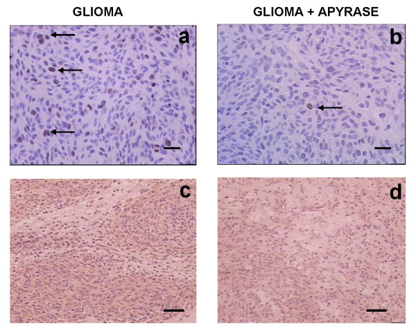
Immunohistochemical stainings of gliomas. Glioma cell proliferation was assessed by immunostaining for Ki67 positive glioma cell nuclei (arrows) in rats implanted with gliomas (a) and in rats co-injected with apyrase (b). The sections were immunostained for VEGF, in rats implanted with gliomas (c) and in rats co-injected with apyrase (d). Scale bars = 20 μm (a,b); 100 μm (c,d).