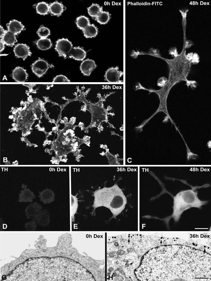
Fig. 1a-h.

Dexamethasone (Dex) treatment induces differentiation of UR61 cells into a sympathetic neuron-like phenotype. a-c Phalloidin-FITC (fluorescein isothiocyanate) was used as marker of cortical actin to demonstrate two progressive stages of neuritogenesis upon dexamethasone treatment for 36 h b and 48 h c. Untreated cells (a) exhibit a round shape with numerous blebs of the plasma membrane. d-f UR61 cells were stained for tyrosine hydroxylase (TH). Weak labeling is detected in the cytoplasm of untreated cells (d), but the cytoplasmic expression of this enzyme dramatically increases upon dexamethasone treatment (e,f). g, h Representative electron micrographs of untreated (g) and dexamethasone-treated (h) UR61 cells. Note in g the narrow band of perinuclear cytoplasm, the absence of secretory granules and the formation of blebs at the cell cortex. Upon dexamethasone-induced differentiation (h) numerous secretory granules (arrows) appear, predominantly distributed in the marginal cytoplasm. Bar represents 5 μm(a-f); 1 μm(g, h)
