
Fig. 5.
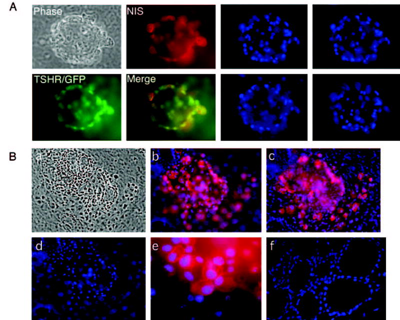
GFP+NIS+ cells are organized into one or more follicle-like cell clusters. Left panel, A follicle-like cluster derived from TSHR+/− ES cells after 21 d of differentiation visualized microscopically (phase) or after staining with an antibody to NIS (red). TSHR expression is indicated by the green GFP signal. An overlaid image shows the colocalization of NIS with TSHR (yellow). Right panel, The same field of cells after nuclear staining with DAPI (blue). Images were taken from different optical sections. B, Immunofluorescent images of several follicle-like clusters derived from TSHR+/− ES cells after 21 d of differentiation. a, Phase contrast exposure. b and c, Immunofluorescent staining, demonstrating the expression of NIS protein. Note that a rim of cells several layers thick adhered closely to the NIS-positive cells. d, Nuclear DAPI staining. e, High magnification of anti-NIS immunofluorescence, demonstrating the expression of NIS in the plasma membrane. Immunofluorescence was not detected when the experiment was performed with the second antibody alone (data not shown). f, Nuclear DAPI staining of native mouse thyroid tissue.