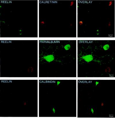
Figure 1.
Confocal microscope images showing the double labeling for Reln and several Ca2+-binding proteins in layers II–III of 20-μm sections of the adult rat frontoparietal cortex. (Top) The double-immunofluorescent labeling for Reln (fluorescein; green) and calretinin (rhodamine; red). (Middle) Double immunolabeling of Reln (gold; red) and parvalbumin (fluorescein; green). (Bottom) The double immunolabeling of Reln (gold; red) and calbindin (fluorescein; green). Overlays (Right) show that Reln and these Ca2+-binding proteins are poorly colocalized in the adult rat frontoparietal cortex.

