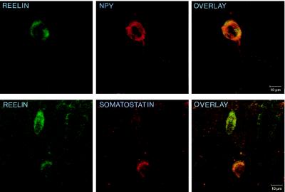
Figure 2.
Confocal microscope images showing the colocalization for Reln and neuropeptides in layers II–III of 20-μm sections of the adult rat frontoparietal cortex. (Upper) Colocalization of Reln (fluorescein; green) and NPY (rhodamine; red). (Lower) Colocalization between Reln (fluorescein; green) and somatostatin (rhodamine; red). Overlays at right are examples of the colocalization frequency between Reln and NPY, or somatostatin reported in Table 1.

