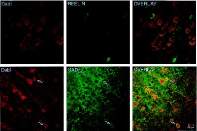
Figure 4.
Confocal microscope images taken from layer V of the adult rat frontoparietal cortex. (Upper) Double labeling of Dab1 (Gold; red) and Reln (fluorescein isothiocyanate; green). Note that Dab1 is predominantly located in pyramidal cells and is not found in Reln-expressing cells. (Lower) Double labeling of Dab1 (Gold; red) and GAD67 (fluorescein isothiocyanate; green). (Left) Whereas Dab1 is predominantly located in pyramidal cells, Dab1 is also found in a small population of nonpyramidal cells (arrows). (Center) Typical distribution pattern of GAD67 (fluorescein; green), both in GABAergic cells (arrows) and in the terminal endings surrounding the excitatory pyramidal cells. The overlay of the two images (Right) shows that these small Dab1-positive cells are GABAergic cells (arrows). Note also that there is a small population of pyramidal cells that do not contain Dab1, evidenced by the GAD67-labeled outline of pyramidal cells that are not Dab1-positive.

