
FIGURE 1.
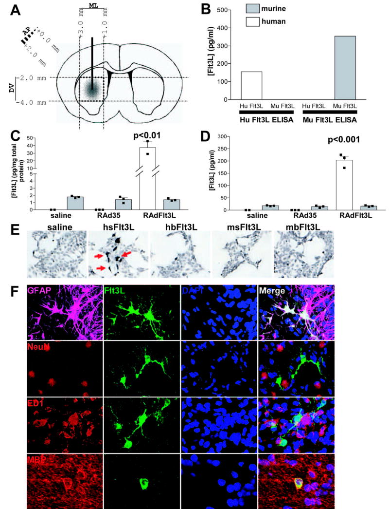
Expression of Flt3L in rat brain using RAdFlt3L. A, Schematic diagram outlining the injection of RAdFlt3L into rat brain striatum and the subsequent dissection of brain tissue around the injection site while carefully avoiding ventricles and meninges. B, Human Flt3L ELISA is specific for recombinant human Flt3L and does not cross-react with recombinant mouse Flt3L. Rodent Flt3L ELISA does not cross-react with recombinant human Flt3L. C, Human Flt3L was elevated 20-fold compared with endogenous rat Flt3L in the brain when animals were injected with RadFlt3L. However, endogenous Flt3L did not change in response to viral injection into the brain. D, Injection of RAdFlt3L in the brain elevated serum concentrations of Flt3L 10-fold over those of endogenous rat serum Flt3L, the concentration of which was not found to change in response to viral injection. E, HEK 293 cells were plated on coverslips in 6-well plates at a density of 1.5 × 10⁵ cells/well. The following day, medium was changed, and cells were transfected with 1 μg of plasmid DNA (pAL119-hsFlt3L, pAL119-hbFlt3L, pAL119-msFlt3L, or pAL119-mbFlt3L) using GeneJuice (Novatech) according to the manufacturer’s instructions. After 48 h, cells were fixed in 4% paraformaldehyde for 5 min and stained for human soluble Flt3L using rabbit anti-Flt3L Ab. Goat anti-rabbit-HRP secondary Ab and 3,3′-diaminobenzidine were used to visualize Flt3L-expressing cells. F, Confocal image showing the expression of human Flt3L (green) and GFAP (magenta), NeuN (red), ED1 (red), or MBP (red), as indicated, within the brain striatum. Double labeling of Flt3L with GFAP- (white) and MBP (yellow)-expressing cells indicates that the majority of RAdFlt3L infected astrocytes and oligodendrocytes. Flt3L was not found to colocalize with NeuN (yellow), suggesting that RAdFlt3L was not expressed in rat neurons. We also observed double labeling of ED1 and RAdFlt3L by confocal microscopy (yellow). However, transverse slices revealed that Flt3L does not colocalize with ED1 and was, instead, indicative of contacts or close association between activated microglia and RAdFlt3L-expressing cells, rather than actual expression of Flt3L by ED1-positive microglia. Results are representative of five animals analyzed in each group.