
FIGURE 7.
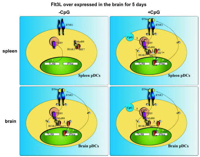
TLR9 signaling in brain- and spleen-derived pDCs. In the absence of CpG, unstimulated pDCs isolated from the spleen (upper left) expressed TLR9 (1). However, IFN-α was not expressed in these cells (A, left). After incubation with CpG (upper right), CpG was endocytosed by spleen pDCs (1) and bound with TLR9 in the endosomal compartment (2). This recruited the TLR9 signaling complex, comprised of MyD88, IRAK1, and IFN-regulatory factor 7 (IRF7), to TLR9. Activation of IRAK1 resulted in IRF7 phosphorylation (3) and translocation to the nucleus (4). IRF7 activates transcription of IFN-α (5), which, in turn, is secreted from the cell and binds with IFNR1 (6). IFNR1 signaling appears to down-regulate TLR9 expression in spleen cells shortly after stimulation with CpG (7). The pDCs isolated from the brain after overexpression of Flt3L (lower left) had lower expression of TLR9 than the naive spleen counterparts. IFN-α was constitutively expressed (3), presumably through MyD88 and IRF7 (1, 2) and was secreted by these cells (4). After incubation with CpG (lower right) (1, 2), IFN-α was not further increased (3), indicating that TLR9 signaling was refractory to superstimulation using CpG.