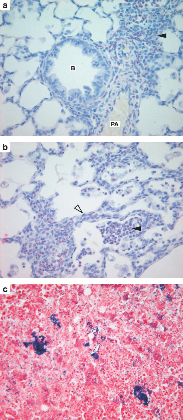
Figure 3. Histology of the Lung and Middle Ear Exudate in Junbo Mice.
(a) 13-d postnatal Jbo/+ lung with perivascular and peribronchiolar cuffs containing Sirius red positive eosinophil leukocytes (arrowhead), bronchiole, pulmonary artery ×400, (b) focal eosiniphilic alveolitis (arrowhead) and thickened alveolar septae (open arrowhead) with eosinophil-rich infiltrates. (c) 21-d postnatal Jbo/+ MEC pus with colonies of Gram positive cocci ×600. B, bronchiole; PA, pulmonary artery

