
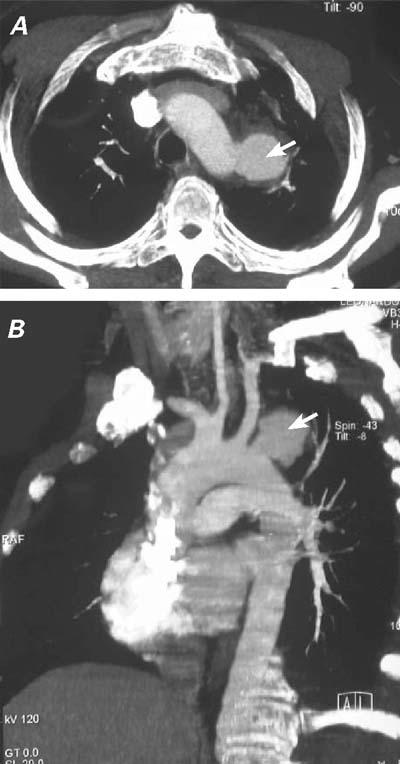
Fig. 2 A) Computed tomographic scan shows the 3.8-cmdiameter aneurysm (arrow) distal to the left subclavian artery. B) Computed tomographic angiography shows the 3.8-cm saccular aneurysm (arrow) with a 1.8-cm neck, located 1 cm below the left subclavian artery. Note that both carotid arteries arise from the brachiocephalic trunk.