
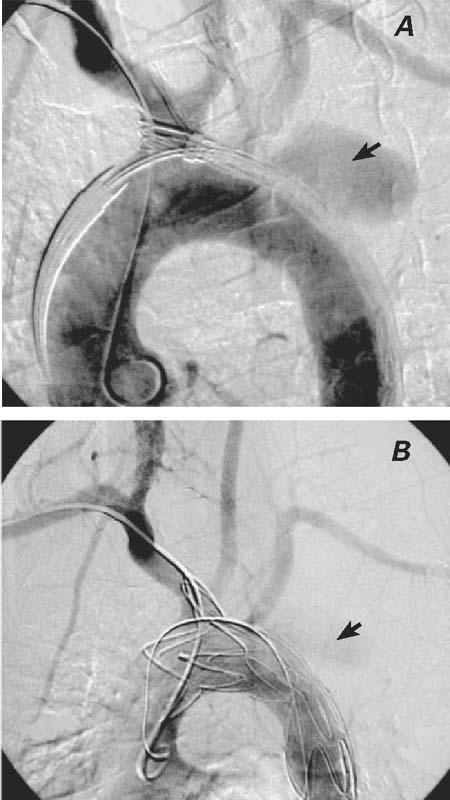
Fig. 3 A) Perioperative angiography shows the positions of the catheters and the unexpanded stent, as well as the brachiocephalic trunk, carotid arteries, subclavian arteries, and the saccular aneurysm (arrow). B) Perioperative angiography shows the successful exclusion of the saccular aneurysm. Left subclavian artery flow from the collateral circulation has not been compromised. There was minor type II endoleakage (arrow) after the stent-graft implantation at the late phase of angiography.