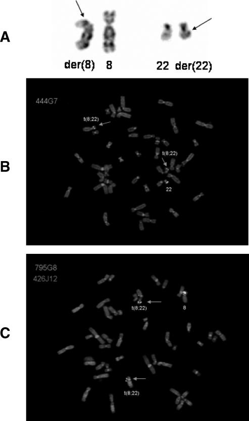
Figure 1.
(A) Partialkaryogram showing a reciprocal translocation t(8;22)(p21; q12). Arrows indicate breakpoints. (B) Metaphase cell hybridized with the BAC probe RP11-444G7 (covering the CHEK2 locus; green signals). Arrows indicate split signals on derivative chromosomes 8 and 22. (C) Metaphase cell hybridized with BAC probes RP11-795G8 (covering the PPP2R2A locus; green signals) and RP11-426J12 (mapping to the centromeric side of the breakpoint on chromosome 8; red signal). Arrows indicate split signals for RP11-795G8 on the derivative chromosomes 8 and 22.

