Figure 4.
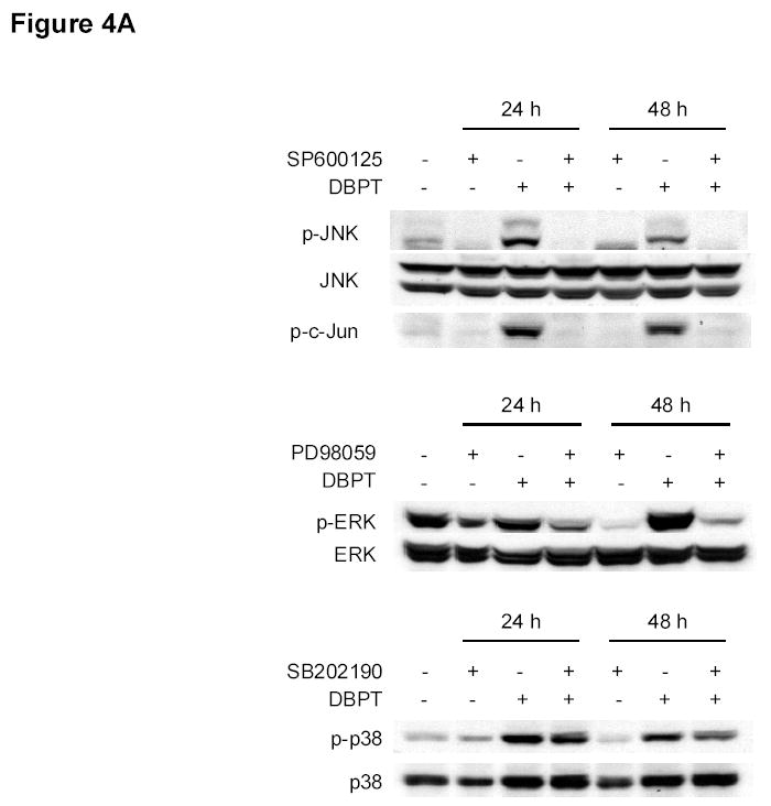
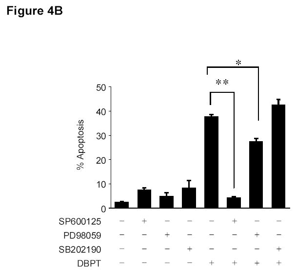
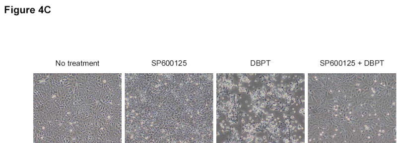
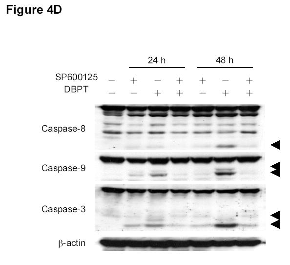
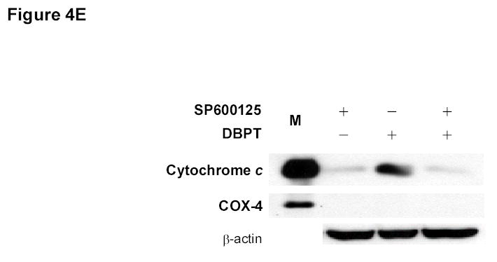
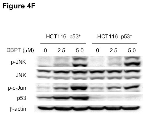
Effect of MAPK inhibitors on DBPT-induced apoptosis. A, DLD-1 cells were treated with 3 μM DBPT in the presence or absence of 50 μM PD98059 (ERK inhibitor), 40 μM SB202190 (p38 inhibitor), or 50 μM SP600125 (JNK inhibitor) for 24 or 48 h. Whole-cell lysates were analyzed for JNK, c-Jun, ERK, and p38 activation by immunoblotting. B, Apoptotic cells after treatment as described in A. Data represent mean ± SD from three independent experiments performed in triplicate. *, P< 0.01, **, P< 0.001 among the indicated groups. C, phase-contrast photomicrographs of DLD-1 cells treated with 50 μM SP600125, 3 μM DBPT or both for 24 h. Magnification, × 100. D, Dection of caspase activation by immunoblotting. DLD-1 cells were treated with DBPT in the presence or absence of SP600125 for 24 or 48 h. Whole-cell lysates were analyzed for caspase activation. β-actin was used as a loading control. Arrowheads indicate cleavage proteins. E, DLD-1 cells were treated with DBPT in the presence or absence of SP600125 for 24 h. Cytosolic fractions were analyzed by immunoblotting with anti-cytochrome c. Mitochondrial fractions (M) were used as a positive control. COX-4 and β-actin were used as a loading controls for mitochondrial and cytosolic fractions. F, wild-type (p53+) and p53-deficient (p53−) HCT116 cells were treated with 2.5 or 5 μM DBPT for 24 h. Cell extracts were analyzed for the indicated proteins by western blotting.
