Figure 5.


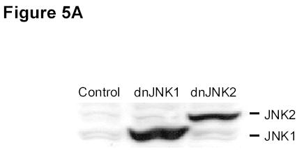
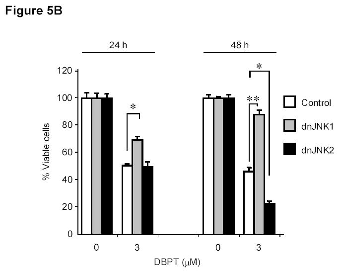
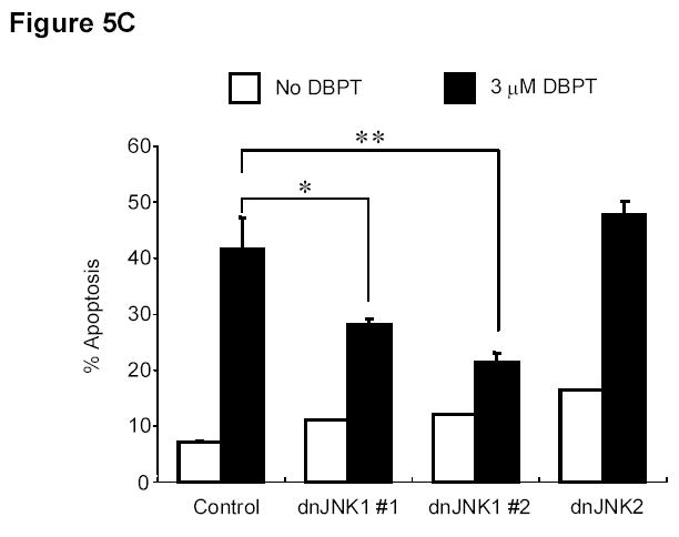
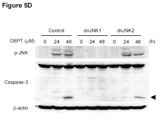
JNK1 activation has a crucial role in DBPT-induced apoptosis. A, DLD-1 cells transfected with dnJNK1 or dnJNK2 plasmids were analyzed by western blot with anti-HA antibodies. DLD-1 cells stably transfected with dnJNK1, dnJNK2 or an empty vector (control) were treated with 3 μM DBPT for 24 or 48 h. Cell viability was determined by XTT assay (B), and apoptotic ratio was determined by flow cytometry (C). Data represent mean ± SD of three independent experiments. *, P< 0.01, **, P< 0.001 among the indicated groups. D, DLD-1 cells stably transfected with dnJNK1 or dnJNK2 were treated with 3 μM DBPT for 24 and 48 h. Whole-cell lysates were analyzed for indicated antibodies by western blot analysis.
