
Figure 4.
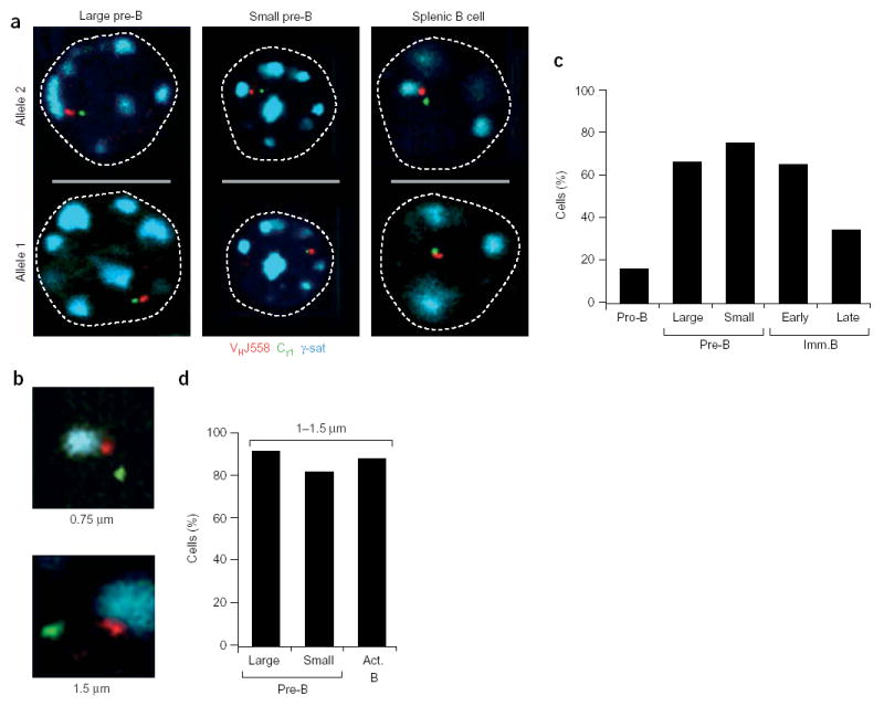
Monoallelic centromeric recruitment of the Igh locus during B cell development. (a) Centromeric location of one Igh allele in bone marrow pre–B cells. Large and small pre–B cells and activated splenic B cells were analyzed by three-color three-dimensional DNA FISH with VHJ558 (red), Cγ1 (green) and γ-satellite (γ-sat; blue) probes. The relative positions of the three signals are shown in confocal sections through the nuclei of these cells. (b) Association of the distal VHJ558 gene domain with centromeric clusters. Enlargements show the orientation and decontraction of the Igh locus at γ-satellite clusters. Below images, distance between the VHJ558 and Cγ1 probe signals. (c) Monoallelic recruitment of the Igh locus to centromeres. Data represent the percentage of cells showing association of one Igh allele with centromeric heterochromatin in various B cell developmental stages, sorted as indicated in Supplementary Fig. 1 online. Actual numbers and sample sizes are in Supplementary Table 2 online. (d) Preferential location of widely separated Igh alleles at the centromeres. The cells showing monoallelic centromeric recruitment were subdivided into a population of cells containing an Igh allele with wide separation (1–1.5 μm) of the VHJ558 and Cγ1 genes. Data represent the percentage of centromeric recruitment of the widely separated Igh allele in this cell population for large and small pre–B cells and activated (Act.) splenic B cells.