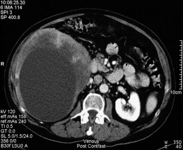
Figure 1.

Portal venous phase image of an axial CT cut showing a large heterogeneous cystic mass within the right lobe of the liver centered on segment 5 and 6 and measuring 15 × 17 × 20 cm in maximum dimension. Further cuts show extension to segment 4A and segment 1 encroachment. No direct portal or peri-coeliac lymphadenopathy was identified.
