
Figure 1.
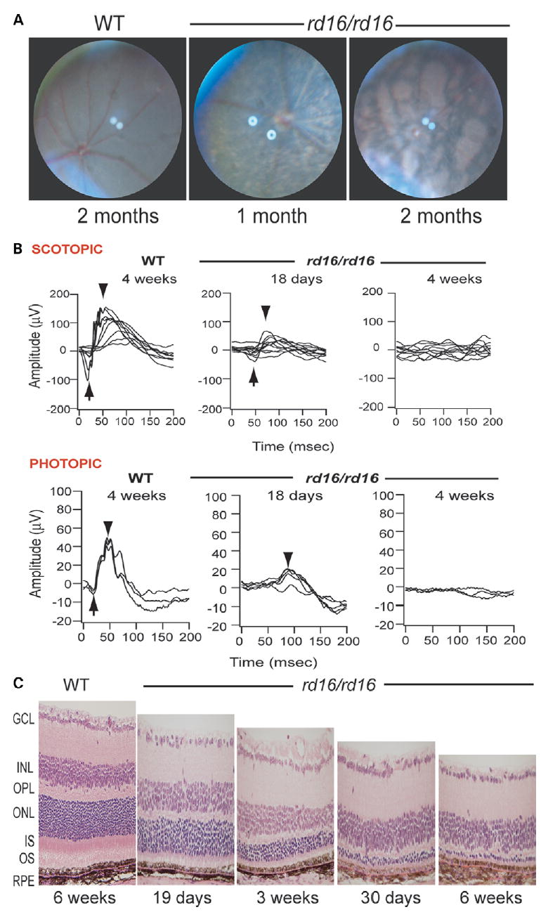
Examination of the rd16 mouse retina. (A) Fundus photographs of WT C57BL/6J mouse and the rd16 homozygote mutants (rd16/rd16) demonstrating retinal degeneration at 1 month of age and at 2 months. (B) ERG responses of WT and mutant (rd16/rd16) mice under dark- (SCOTOPIC) and light- (PHOTOPIC) adapted conditions. Arrows indicate the A-wave and arrowheads the B-wave. (C) Histology of retina ofWT and rd16 homozygotesmice at indicated ages. RPE, retinal pigment epithelium; OS, outer segments; IS, inner segments; ONL, outer nuclear layer; OPL, outer plexiformlayer; INL, inner nuclear layer; GCL, ganglion cell layer.