
Figure 5.
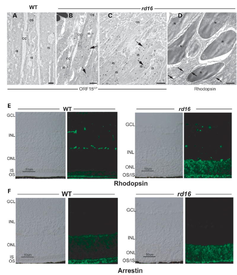
Localization of RPGR-ORF15, rhodopsin and arrestin in rd16 retinas. (A–D) Immunogold EM of WT or rd16 retinas with indicated antibodies. Labeling with ORF15CP antibody showed a predominant connecting cilium (CC) staining of RPGR-ORF15 (A) as opposed to abnormal extensive labeling throughout the photoreceptor IS in the rd16 retina (B, C). Arrows indicate clusters of immunogold particles. Labeling of rhodopsin in the rd16 retina (D) is evident around the photoreceptor cell bodies (indicated by arrows) with no exclusive OS localization; N, nucleus. (E, F) Immunohistochemical analysis of the WT and rd16 retinas at P12, dissected under normal light/dark cycle, with antibodies against rhodopsin (E) or arrestin (F). As shown, both rhodopsin and arrestin are localized primarily in the OS of WT retina, whereas in rd16, rhodopsin and arrestin are also detected in the ONL and ISs of photoreceptors. OS in the rd16 retina degenerate at P12 and therefore are represented in conjunction with the inner segments (OS/IS). Scale bar: 50 μm.