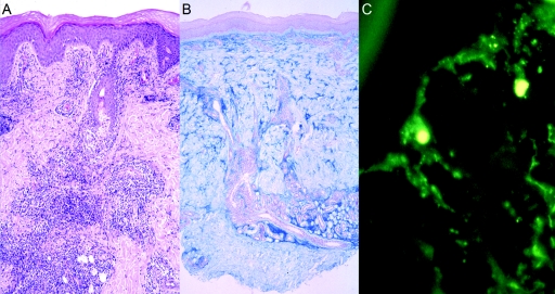
Figure 3. .
Histology of lesional skin biopsy from proband V1. A, Hematoxylin-eosin staining (magnification 1:100) showing lamellar orthohyperkeratosis and regions of hydropic degeneration of the stratum basale as well as single-cell necrosis. Inflammatory infiltrates with perivascular and periadnexial distribution and interface dermatitis can be seen. B, Alcian blue staining (magnification 1:40) showing increased deposits of mucin throughout entire stratum reticulare. C, Direct immune fluorescence (magnification 1:100) showing broad granular deposits of C3 along basement membrane zone. A similar pattern was also seen with staining for IgM or IgA.

