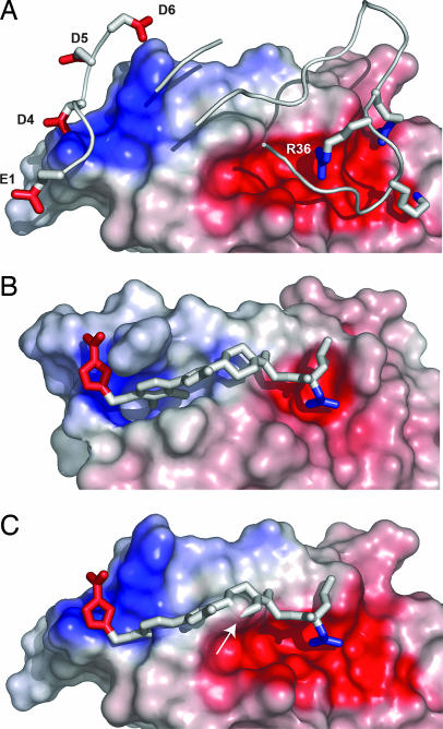
Fig. 3.
Binding surfaces and electrostatic fields created by IL-2. (A) IL-2Rα in ribbons and sticks with specific residues labeled binding to the surface of IL-2, which is colored in proportion to the electrostatic field that would be felt by a test atom in contact with the surface. Blue is positive, white is neutral, and red is negative, with a dynamic range of −15 to +15 kBT/ec at T = 310 K, and all potentials are calculated with respect to the dielectric envelope of the fully solvated complex. (B) SP4206 in sticks binding to IL-2 represented as in A. Note the more localized electrostatic field presented by IL-2 in the presence of SP4206. (C) Bound conformation of SP4206 superimposed on the surface of IL-2 that binds the IL-2Rα. Note that in this conformation part of the surface of IL-2 (shown by the arrow) is incompatible with binding.

