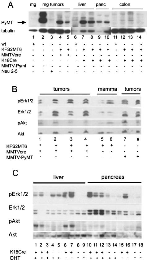
Figure 7.
A: PyMT protein expression in tumors. PyMT and β-tubulin were detected by Western blot analysis. The filter was first reacted with PyMT antibody then stripped and reprobed for tubulin. Twenty micrograms of protein was loaded in each lane except for lane 2, which received 2 μg. The tubulin exposure of lanes 6 to 10 was 5 minutes, whereas the remainder of the image represents a 0.5-minute exposure of the same filter. The genotypes of the animals from which the tissues were derived are shown at the bottom. Neu 2–5, represents line MMTV-Neundl2–5.28 mg, mammary gland; panc, pancreas. B: Frozen mammary tumors and normal whole mammary fat pad (mamma) were subjected to Western blot analysis. Genotype of the tissues is indicated at the bottom. Each lane contains tissue or tumor protein from a different animal. pErk1/2, phospho-Erk1 and phospho-Erk2. pAkt, phosphorylated form of Akt. C: Liver and pancreatic tissues from KFS2MT6 mice with additional genes or treatment indicated below lane numbers. K18Cre indicates presence of K18CreER transgene. OHT indicates 5-day treatment with 5-hydroxy-tamoxifen. Note increased pAkt in bigenic KFS2MT6; K18Cre liver (lanes 1, 2, 6, and 7) and increase pErk1/2 in bigenic KFS2MT6; K18CreER in pancreas (lanes 10 to 12).

