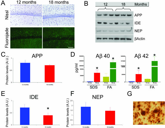
Figure 3.
Investigation of experimental parameters that could underlie the age-related decrease in intraneuronal Aβ. A: Representative microphotographs of sections stained with the Nissl and fluorojade technique indicating that there is no evident loss of neurons between 12 and 18 months of age (n = 6 per group). B: Representative immunoblots of protein extracts from the hippocampus of 12- and 18-month-old 3xTg-AD mice analyzed for APP, IDE, and NEP. β-Actin levels were assayed to control for protein loading. Note that the representative blots are n = 3 whereas the quantifications of these blots (see below) are done one a sample of n = 6. C: Quantitative analysis of APP blots shows that full-length APP steady-state levels remain relatively constant during these time points (P = 0.158). D: ELISA measurement of Aβ40 and Aβ42 in 12- and 18-month-old 3xTg-AD mice. There is a significant age-dependent increase in both Aβ40 (P = 0.0054 and P < 0.0001 for sodium dodecyl sulfate and FA, respectively) and Aβ42 levels (P = 0.0476 and P = 0.0038 for sodium dodecyl sulfate and FA, respectively), between these time points. E: Steady-state levels of IDE show a significant age-dependent decrease (P < 0.05). F: A similar decrease was also detected for NEP, although the difference did not achieve statistical significance (P > 0.05). G: Representative microphotograph showing staining of extracellular plaques and nearby neurons that contain intraneuronal Aβ immunoreactivity. Scale bars: 125 μm (A); 50 μm (G).

