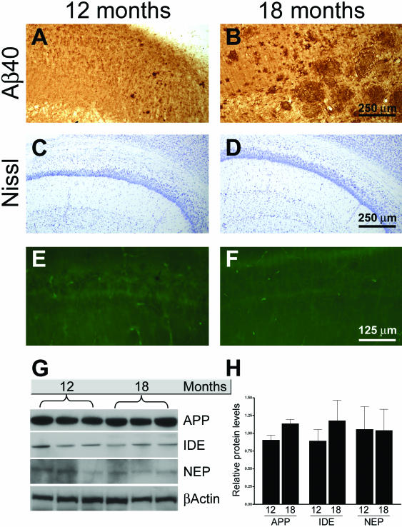
Figure 4.
Intraneuronal Aβ immunoreactivity decreases as a function of age in Tg2576 mice. A and B: Sections from 12- and 18-month-old mice (n = 5) were stained with an Aβ40-specific antibody. The photomicrographs reveal intraneuronal Aβ immunoreactivity in 12-month-old mice, which is absent in 18-month-old mice. In contrary, the number of plaques greatly increases during this time. C–F: Representative microphotographs of hippocampal sections stained with the Nissl and fluorojade, respectively, showing that there is no evident loss of neurons between 12 and 18 months of age. G: Representative Western blots showing APP, IDE, and NEP levels as a function of age. H: Quantification of Western blots for APP, IDE, and NEP show no significant changes in the steady-state levels of these proteins between 12- and 18-month old mice. The levels of each time point were adjusted to the relative β-actin levels, which were used as a protein loading control.

