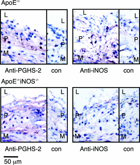
Figure 2.
A comparison of PGHS-2 and iNOS localization in aortic sinus lesions from ApoE−/−and ApoE−/−iNOS−/−mice. Immunohistochemical detection of iNOS and PGHS-2 in aortic sinus lesions from ApoE−/−(top) and ApoE−/−iNOS−/− (bottom) mice fed a Western diet for 4 months. L, lumen; P, plaque; M, media; con, control sections stained with antibody preincubated with antigen. Sections were counterstained with hematoxylin to visualize nuclei. Similar results were observed in aortic sinus lesions from three ApoE−/− and three ApoE−/−iNOS−/− mice.

