Figure 5.
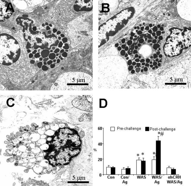
Mast cell activation. Jejunal tissues were obtained from naive control rats (Con), stressed (WAS/Ag) rats, sham-stressed (Con/Ag) rats, WAS-only (WAS) rats, and WAS rats pretreated with the CRH antagonist, α-hCRH (αhCRH/WAS/Ag). All rats except naive controls received a bolus of intragastric HRP at day 5 of the stress/sham protocol; 15 days later tissues were obtained and mounted in Ussing chambers. Ninety minutes after HRP antigen challenge, tissues were removed and processed for electron microscopy. A–C: Electron photomicrographs show mucosal mast cells in the lamina propria. A and B: Representative sections from a Con/Ag rat (A) and rat treated with α-hCRH (B) show normal mast cells with electron dense granules. C: Representative section from a WAS/Ag rat shows a mast cell with depleted granules. D: Percentage of mast cells degranulated after HRP antigen challenge as measured by empty granules or reduced density of granules. Values indicate mean ± SEM; n = 6 rats in each group (20 random views per rat section averaged to obtain each rat value). *P < 0.05, compared with Con group; #P < 0.05, compared with WAS-only group.

