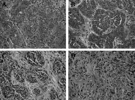
Figure 1.
Histological appearance of the C4-HD tumor during growth and regression. BALB/c mice bearing the C4-HD tumor were treated by removing MPA or by adding estradiol (E2) or the anti-progestin RU486 for 12, 24, 48, or 72 hours to induce tumor regression. Tumors were collected, fixed in 10% formalin, embedded in paraffin, and processed according to standard procedures to obtain H&E-stained sections. A: Appearance of the growing C4-HD tumor (control) is characterized by groups of atypical cells with cribriform differentiation and occasional central necrosis. Stroma is fibroblastic and well vascularized. Atypical features, as well as several mitotic figures, are evident at high magnification. Representative H&E sections prepared from tumors derived from mice treated with RU486 for 12 (B), 24 (C), and 72 (D) hours are shown. Similar images were observed in the −MPA- and E2-treated samples. B: At 12 hours the tumor shows round groups of cells with extensive apoptosis or central necrosis. The stroma is more abundant as the intercellular substance seems to have increased. C: By 24 hours only a few tumor cells appear viable. Many of them have undergone apoptosis or are part of a necrotic process. Stroma is very abundant and seems well vascularized. D: At 72 hours few viable tumor cells are evident. Cellular debris and apoptotic fragments are embedded in an abundant fibroblastic stroma. Original magnifications, ×400.

