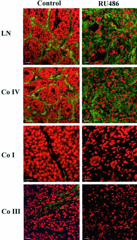
Figure 4.
Remodeling of ECM components during tumor regression. Mice bearing the C4-HD tumor were treated for 72 hours by either removing MPA or adding E2 or RU486 to induce regression. Samples were collected, frozen, and processed for immunofluorescence. Nuclei were stained with propidium iodide (red) and ECM components with the corresponding primary antibodies followed by FITC-conjugated secondary antibodies (green). The panels show staining for laminin (LN), collagen IV (Co IV), collagen I (Co I), and collagen III (Co III) in samples derived from control and 72-hour-treated RU486 mice. Note the change in ECM composition in the treated samples. The same pattern of staining was detected in tumors derived from animals treated by only removing MPA or by adding E2 (not shown). Scale bars, 20 μm.

