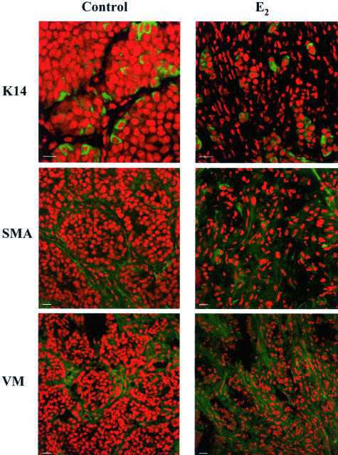
Figure 5.
Characterization of cell types involved in the stromal reaction during regression. Tumors from control, and −MPA-, E2-, and RU486-treated mice were collected at 72 hours. Samples were frozen and processed for immunofluorescence. Nuclei were stained with propidium iodide (red) and cytokeratin 14 (K14), α-smooth muscle actin (SMA), and vimentin (VM) were detected using the corresponding primary antibodies followed by FITC-conjugated secondary antibodies (green). The samples shown correspond to 72-hour E2-treated samples. Similar results were obtained from samples derived from −MPA- and RU486-treated mice. An increase in VM- and SMA-positive cells was observed. K14-positive cells remained surrounded by fibroblast cells that were negative for this myoepithelial cell marker. Scale bars, 20 μm.

