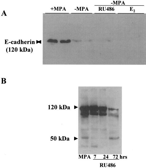
Figure 8.
Fragmentation of E-cadherin during tumor regression. Tumors from control and −MPA-, E2-, and RU486-treated mice were collected at 7, 24, and 72 hours. A: Western blot in which control samples were run together with samples derived from mice treated for 72 hours by either removing MPA (−MPA) or adding E2 or RU486. The Santa Cruz antibody was used in this case. A 120-kd band corresponding to the molecular weight of E-cadherin was detected in the control samples. On 72 hours of regression, the band was practically undetectable. B: Samples from control and 7-, 24-, and 72-hour RU486-treated mice run on an 8% gel. In this case the Transduction Laboratories antibody was used. Several isoforms of E-cadherin were detected using this antibody in control mice. On regression the high-molecular weight bands were reduced considerably and the 50-kd and other small molecular weight fragments were increased. Similar results were obtained with samples derived from −MPA- and E2 treated mice (not shown).

