Figure 5.
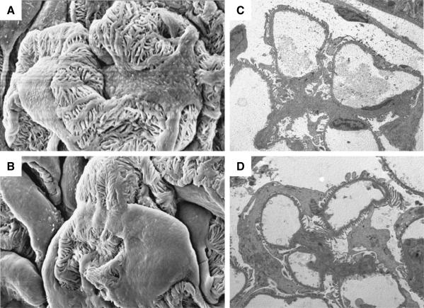
Representative SEM (A and B; final magnification, ×3000) and TEM (C and D; final magnification, ×2200) micrographs of podocyte ultrastructure in Wistar (A and C) and MWF rats (B and D) of 20 weeks of age. Podocyte body covers most of the peripheral capillary wall surface in MWF animals (B and D).

