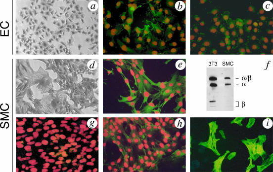
Figure 1.
Expression of HGF/SF and MET in ECs and SMCs. Phase contrast image (a) and expression of HGF/SF (b) or MET (c) by immunofluorescence in subconfluent cultures of rat ECs. Phase contrast image (d) and expression of HGF/SF in SMCs by immunofluorescence (e) or Western blot (f). Both ECs (b) and SMCs (e) express HGF/SF but expression in ECs is weaker and not homogenous; in contrast, SMCs show strong and homogeneous expression of HGF/SF. f: The HGF/SF protein secreted by SMC cultures is indistinguishable from the protein isolated from a 3T3 line.14 g and h: Expression of MET in SMCs. Confluent cultures of SMCs do not express MET (g) but SMCs migrating at the edge of a scrape injury (as shown in Figure 2) show clear, positive staining for MET (h). i: MET-expressing cells from SMC cultures are positive for α-smooth muscle actin. Immunofluorescence of HGF/SF or MET is demonstrated by green fluorescence with nuclear counterstain in red.

