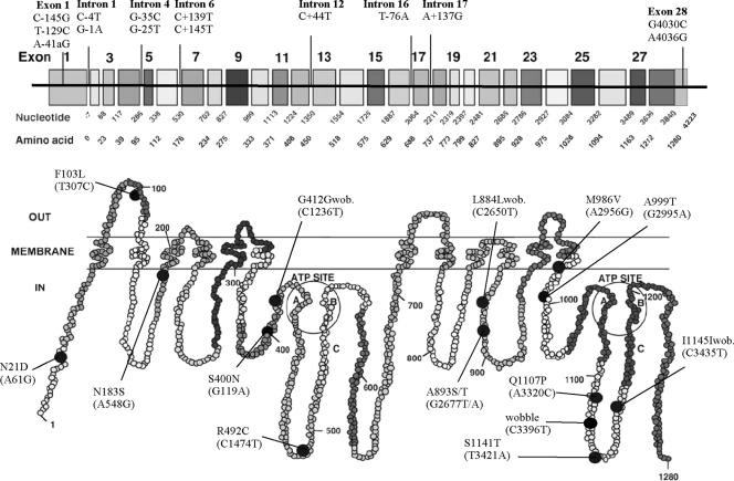
FIG. 3.
Two-dimensional (2D) structure of P-gp showing the distribution of coding SNPs (black-filled circles) (229). The amino acid substitutions are presented, together with nucleotide changes (in parentheses). At the top, 28 exons of the MDR1 (ABCB1) gene are shown with the cDNA nucleotide and amino acid positions, together with noncoding SNPs at the promoter, introns, and untranslated regions. The region of P-gp encoded by a given exon is also highlighted in similar shading on the predicted 2D P-gp structure. The positions of the polymorphisms correspond to the positions of MDR1 (ABCB1) cDNA, with the first base of the ATG start codon set to 1. Mutations located in introns are given as the position downstream (−) or upstream (+) of the respective exon according to the genomic organization of MDR1 (ABCB1) (41). wob., wobble. (Modified from reference 3 by permission from Macmillan Publishers Ltd.)

