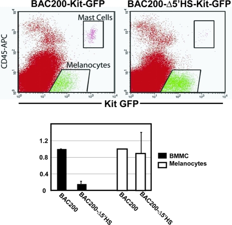
FIG. 3.
Kit-GFP reporter expression in mast cells and melanocytes in P4 back skin of BAC200-Kit-GFP (T20) and BAC200-Δ5′HS-Kit-GFP transgenic mice (T92). Cell suspensions of dorsal skin were stained with APC-conjugated anti-Kit and anti-CD45 antibodies, and cells were analyzed by FACS on the basis of Kit, CD45, and GFP expression. In the bar graph, the numbers of Kit+ CD45+ (mast cell subset) and Kit+ CD45− (melanocyte subset) cells expressing Kit-GFP from BAC200-Δ5′HS-Kit-GFP mice relative to the numbers of Kit-GFP expressing Kit+ CD45+ and Kit+ CD45− cells from BAC200-Kit-GFP mice are shown. The standard deviations are indicated by the error bars (n = 3).

