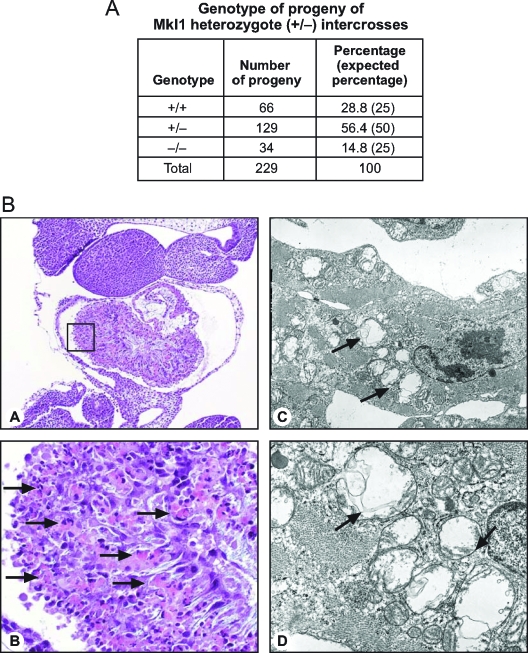
FIG. 2.
Partial embryonic lethality associated with abnormal cardiogenesis due to Mkl1 absence. (A) Mendelian frequency of the progeny born to Mkl1 hemizygote intercrosses. Note that only 14.8% of the offspring are homozygous Mkl1−/− mice (34 of a total of 229 pups), which is less than the expected frequency (25%), indicating partial embryonic lethality in embryos that lack Mkl1. (B) Cardiac sections of E10.5 Mkl1−/− mouse embryos. (A and B) Hematoxylin and eosin staining. (A) Magnification, 40×. (B) Magnification (400×) of boxed area in panel A. There is substantial necrosis of myocardial cells of the ventricular and atrial walls, which appear as hyalinized eosinophilic cells with shrunken and fragmented nuclei (arrows). (C and D) Electron microscopy of E10.5 Mkl1−/− embryonic heart. Mitochondria (arrows) of the affected myocardiocytes are markedly swollen and are shown in various stages of degeneration. There is no cytosolic swelling or change in nuclear morphology suggestive of an apoptotic cell death process; similarly, no electron microscopic evidence of autophagic death (i.e., double-membraned autophagosomes or autophagolysosomes in the cytoplasm) was observed.

