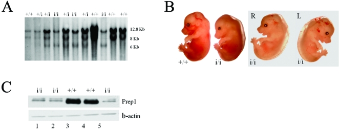
FIG. 1.
Prep1i/i phenotype. (A) Southern blotting analysis of EcoRI-digested DNAs from the progeny obtained by crossing F1 Prep1i/+ × Prep1i/+. (B) Gross morphology of Prep1i/i embryos. The two rightmost panels show the same embryo viewed from both sides (R and L), exhibiting edema, pallor, smaller size, small liver spot and hemorrhaging. (C) Nuclear extracts prepared from E14.5 embryonic brains of 5 littermate embryos (2 wt and 3 Prep1i/i, as indicated) were immunoblotted with monoclonal anti-Prep1 and anti-beta-actin antibodies. Lane 1, 2, and 5 are extracts from Prep1i/i embryos; lanes 3 and 4 are extracts from wt embryos.

