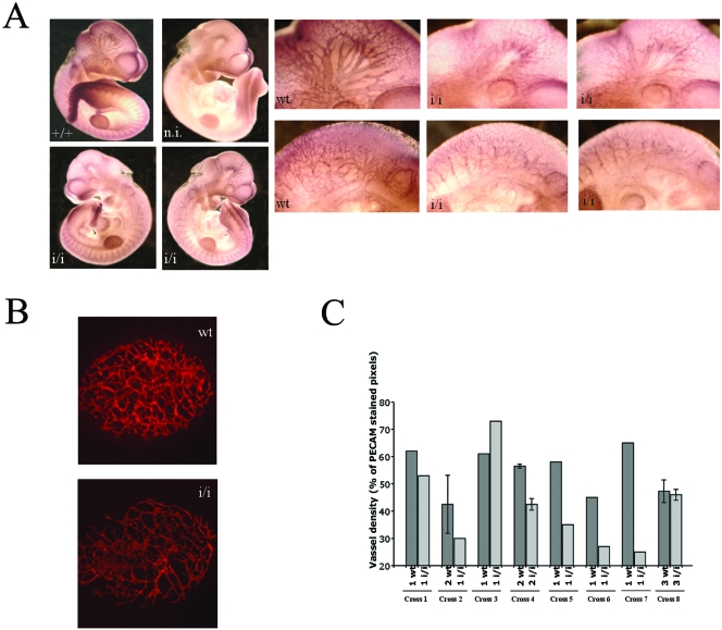
FIG. 3.
Prep1i/i embryos exhibit angiogenesis defects. (A) Whole-mount CD31 (PECAM) immunofluorescence on E10.5 embryos. The genotype is shown in each panel. A nonimmune (n.i.) serum gave essentially no staining (not shown). A set of close-up pictures is inserted. The top row shows details of the head region; the bottom row the intersomitic area. (B) Immunofluorescence on E7.5 wt and Prep1i/i allantois cultured for 18 h and stained with anti-CD31 antibodies. (C) Vessel density (percentage of CD31-stained pixels) in 11 different litters containing wt and Prep1i/i embryos. At the bottom of each histogram, the symbols indicate the numbers of wt and Prep1i/i littermates from different crosses.

