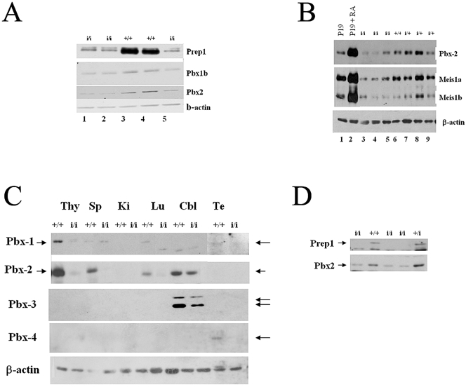
FIG. 7.
Prep1i/i embryonic and adult organs show a decrease of Pbx1b, Pbx2, and Meis1 proteins. (A) Immunoblotting analysis of the same filter shown in Fig. 1C: nuclear extracts from the E14.5 embryonic brain of 5 littermate embryos (2 wt and 3 Prep1i/i, as indicated) tested with monoclonal anti-Pbx1b, anti-Pbx2, and anti-beta-actin antibodies. Lanes 1, 2, and 5 contain extracts from Prep1i/i embryos; lanes 3 and 4 contain extracts from wt embryos. (B) Immunoblotting analysis of brain extracts from 1 wt, 3 heterozygous, and 3 Prep1i/i embryos, using anti-Pbx2 and anti-Meis1 antibodies. (C) Immunoblotting analysis of nuclear extracts obtained from organs, indicated at the top of the panel, of an adult Prep1i/i mouse, tested with anti-Pbx1, anti-Pbx2, anti-Pbx3, anti-Pbx4, and antiactin antibodies. Cbl, cerebellum; Lu, lung; Ki, kidney; Sp, spleen; Te, testis; Thy, thymus. (D) Immunoblotting analysis of E14.5 liver extracts from 1 wt, 1 heterozygous, and 3 Prep1i/i embryos, using anti-Pbx2 (α-Pbx2) and anti-Prep1 (α-Prep1) antibodies.

