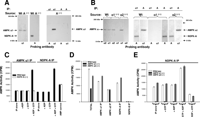
FIG. 1.
Investigation of AMPK α1, AMPK α2, and NDPK-A knockout tissues. The precipitating tissue source is shown above each blot together (where relevant) with the precipitating antibody. The lower part of each blot denotes a 1/1,000 dilution of probing antibody as indicated. (A) Ten micrograms of extracted NDPK-A wild-type (Wt) or NDPK-A-null (−/−) cytosol probed with sheep anti-AMPK α1 or mouse anti-NDPK-A (left and middle panels). The right panel shows anti-AMPK α1 or mouse anti-NDPK-A antibodies used to precipitate (IP) 10 μg of extracted cytosol and probed with either an AMPK α1- or NDPK-A-specific antibody, as indicated. (B) The procedure used was the same as that described above (A), except that the tissue source was extracted liver cytosol from murine wild-type (Wt), AMPK α1 knockout (−/−), or AMPK α2 knockout (−/−) tissue. (C) Ten micrograms of extracted NDPK-A wild-type or NDPK-A-null (−/−) cytosol precipitated using either an AMPK α1- or NDPK-A specific antibody and assayed for AMPK-SAMS activity in the presence/absence of 500 nM ADP, GTP, and ADP plus GTP, as indicated. Error bars (n = 3) on all assay histograms indicate the ranges and not the standard errors. (D) Ten micrograms of extracted AMPK wild-type, AMPK α1-null (−/−), or AMPK α2-null (−/−) cytosol precipitated using either an AMPK α1-, AMPK α2-, NDPK-A-, or NDPK-B-specific antibody and assayed for AMPK-SAMS activity relative to the input (total). (E) Ten micrograms of extracted AMPK wild-type, AMPK α1-null (−/−), or AMPK α2-null (−/−) cytosol precipitated using an NDPK-A-specific antibody and assayed for AMPK-SAMS activity in the presence/absence of 500 nM ADP, GTP, and ADP plus GTP, as indicated; the middle bars show no activity in each triplet.

